 Multiple Sclerosis (MS) is an autoimmune inflammatory disease that affects nearly 2.3 million young adults worldwide. In cases of MS, the immune system promotes an attack on the central nervous system (CNS), often leading to disability and degeneration.
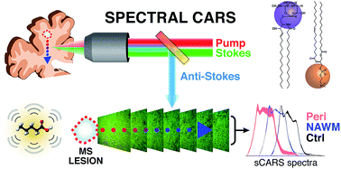
The MS lesion is traditionally considered the leading indicator of CNS damage and thus has been studied for decades through various clinical pathological methods. However, it has been found that surrounding regions in the brain, known as ‘normal-appearing’ white matter (NAWM), also present some abnormalities in MS cases.
Label-free imaging techniques, such as coherent anti-Stokes Raman scattering (CARS) and Stimulated Raman scattering (SRS) have proven to be effective tools for investigating these NAWM abnormalities due to their ability to accurately examine lipid-rich structures like myelin. Prof. Peter Stys and his research group at the Hotchkiss Brain Institute at the University of Calgary studied these abnormalities in the NAWM regions with these methods: in their research, they utilized Clark-MXR’s IMPULSE fiber laser coupled with novel dual-NOPA setup to perform spectrally chirped CARS (sCARS).
Read more: Further information can be found in “Lipid biochemical changes detected in normal appearing white matter of chronic multiple sclerosis by spectral coherent Raman imaging”, K. W. C. Poon, C. Brideau, R. Klaver, G. J. Schenk, J. J. Geurts and P. K. Stys. Chem. Sci., 2018, Advance Article, DOI: 10.1039/C7SC03992A